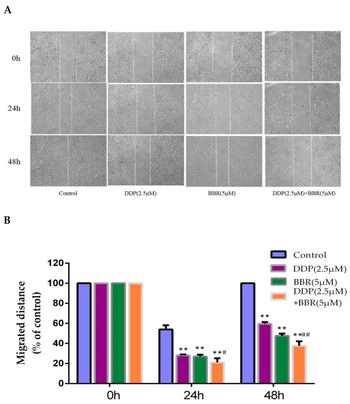
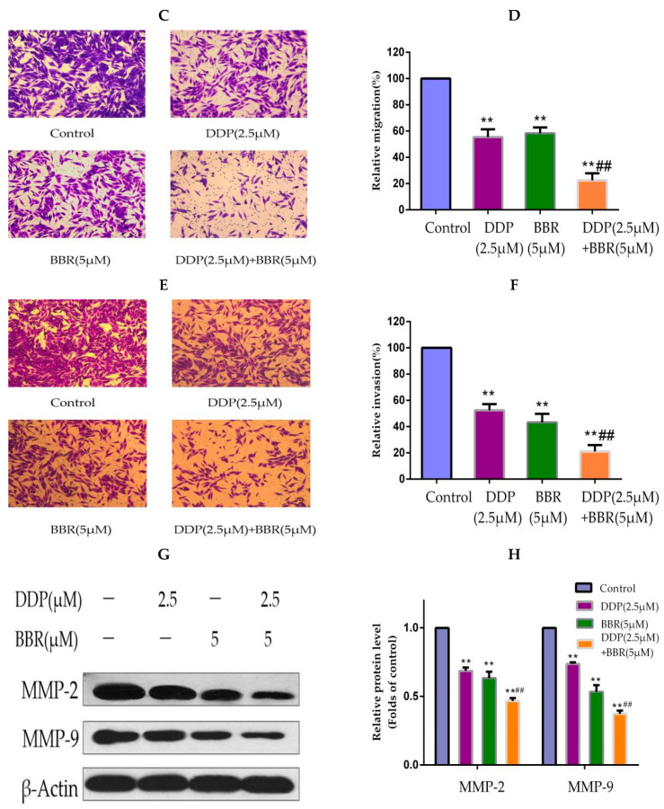
Figure 2.
BBR and DDP suppress the migration and invasion capacity of MG-63 cells. (A,B) Micrographs of wound-healing assays with MG-63 cells treated with BBR and/or DDP. Images were obtained at 0, 24, and 48 h (100× magnification). (C,D) A transwell assay was used to detect the migration of MG-63 cells. The number of migratory cells was observed and counted by using a light microscope (100× magnification). (E,F) A transwell assay was used to detect the invasion of MG-63 cells. The number of invasive cells was observed and counted by using a light microscope (100× magnification). (G,H) Western blot analysis of MMP-2 and MMP-9 in MG-63 cells. The data are presented as the mean ± SD of three independent experiments; ** p < 0.01, compared with the control group. ## p < 0.01, compared with the monotherapy group.


