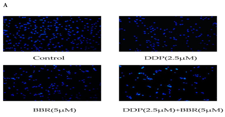
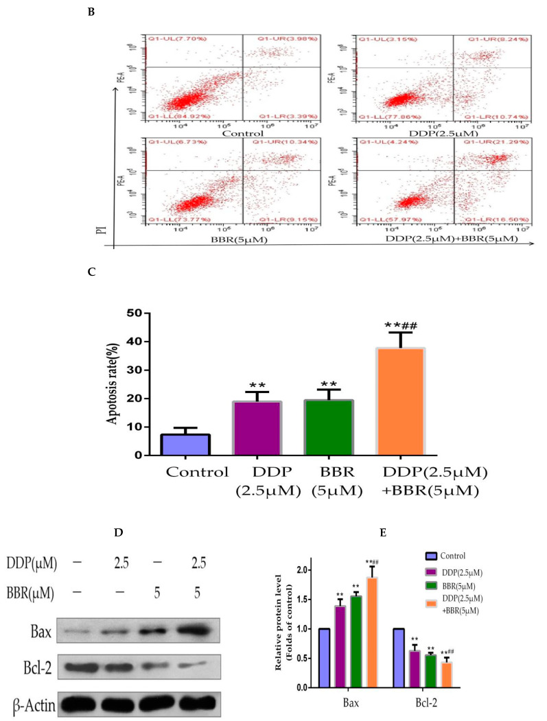
Figure 4.
Effect of BBR and DDP alone and in combination on apoptosis. (A) Hoechst 33258 staining of MG-63 cells treated with BBR and/or DDP for 48 h. Apoptotic cells were identified by the presence of bright-blue fluorescence and highly condensed or fragmented nuclei (100× magnification). (B,C) The apoptosis of MG-63 cells was determined by flow cytometry after staining with annexin V-FITC/P. (D,E) The levels of cleaved Bcl-2 (antiapoptotic protein) and Bax (proapoptotic protein) were detected by Western blotting. The data are presented as the mean ± SD of three independent experiments; ** p < 0.01, compared with the control group. ## p < 0.01, compared with the monotherapy group.


