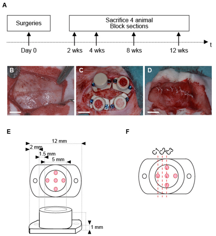
Figure 1.
(A) Time frame of the study. (B–D) Main stages of the surgical procedure: (B) exposition of the calvarium, note the bone sutures that delineate 4 quarters; (C) placement and filling of the cylinders in the 4 quarters delimited by the bone sutures; (D) site suture; (white bars, 5mm); (E) Cylinder specification. Red circles mark the position of the intramedullary holes drilled on rabbit skulls; (F) Schema showing the three different levels of cut (C1–3) within the sample for histological and histomorphometric analyses.

