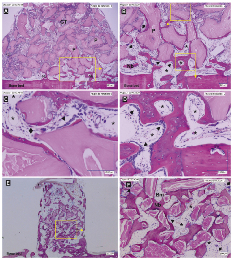
Figure 4.
Representative histological slides showing the entire content of a cylinder filled with a DBBM-C scaffold pre-treated with NaCl 0.9% at 2 weeks (A) and 12 weeks (E) and higher magnifications ((B–D): 2 wks; (F): 12 weeks). Hematoxylin-eosin staining, particles (P) appears as pink, Bone and New bone (Nb) appear as purple. (GT: granulation tissue; Dark arrows ►: lines of osteoblast; Bm: bone marrow; *: capillaries; ♦: osteoclasts; Yellow boxes and arrows: magnifications on the pictures indicated by the arrows.) (A) At 2 weeks, the new bone sprouts from the bone bed and the transcortical holes. Osteoid and new bone tissue are observed in close vicinity to the bone bed. The granulation tissue has already migrated to the top of the scaffold. Below the GT is the osteogenic zone (B,C), a highly vascularized tissue where new bone is deposited at the mineral particle surface. Note the lines of osteoblasts that deposit layers of new bone around the particles (B,C). Some osteoclasts are also observed at the surface of the particles not yet osseointegrated, followed by osteoblast to form basic multicellular units (C). In close vicinity to the bone bed, the remodeling zone is found formed by new bone trabeculae and a vessel rich connective tissue (D). At 12 weeks, all the particles are osseointegrated and the bone has filled the whole cylinder (E). Note the signs of bone maturation with the presence of bone marrow foci in the bone lacunae as well as the presence of lamellar bone (F).

