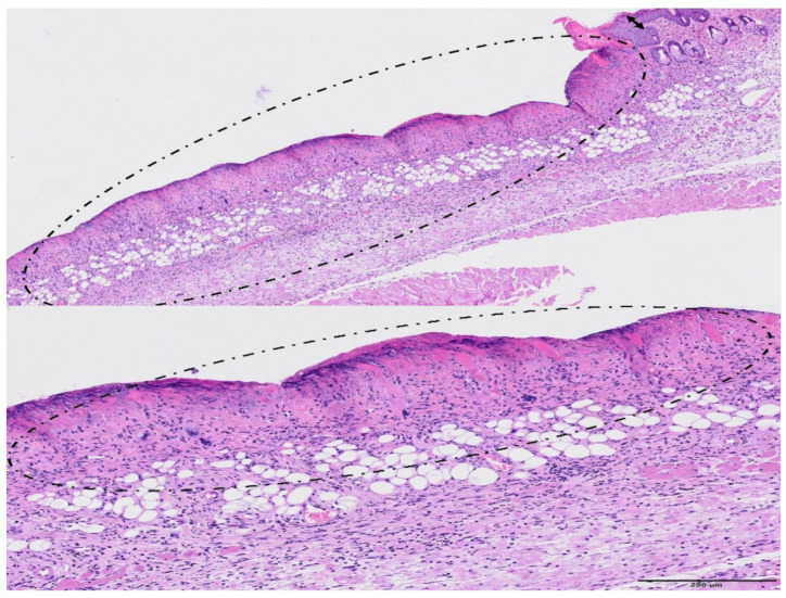
Figure 11.
Broad beam irradiation. HE staining. Upper figure (bar = 500 µm): a large area of the skin presented extensive epidermal necrosis (encircled), with abundant inflammatory infiltration and loss of annexa. There was also epidermal hyperplasia in less affected areas (double-headed arrow). Lower figure—Bar = 200 µm: note that the loss of cellular detail of the epidermis (epidermal necrosis, encircled) at higher magnification. The subjacent dermis appears highly infiltrated by inflammatory cells.

