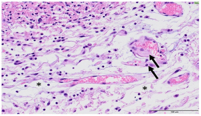
Figure 12.
Broad beam irradiation. HE staining, Bar = 100 µm. Within the dermis, there is an inflammatory infiltrate consisting of macrophages, lymphocytes, and neutrophils. The collagen fibres from the connective tissue were separated by oedema (white spaces, asterisk). In the image, a couple of vessels present early hyalinization (arrows).

