Figure 7.
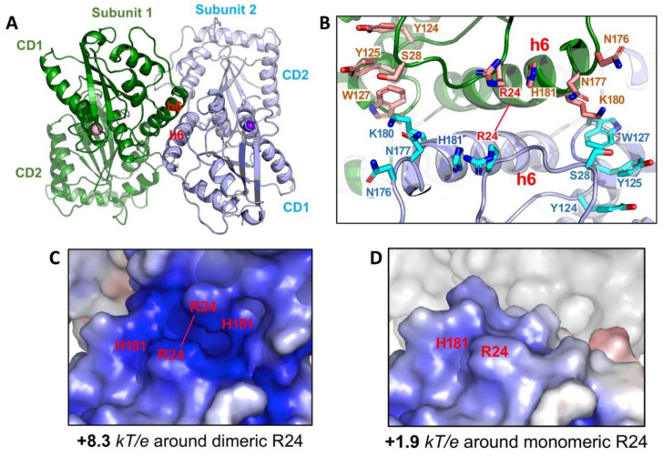
Structure of full-length rA3G. (A) The rA3G dimer structure (6P3X). Dimerization is mediated through CD1-CD1 contacts centered around helix 6 (h6) of CD1. (B) The positively charged residues centered around R24 on both sides of the dimer junction. Dimerization brings these positively charged residues from each subunit to close proximity. (C) The closely positioned charged residues around the dimer junction result in significantly enhanced positively charged electrostatic potentials (PEP or diPEP), as shown by the blue surface (+8.3 kT/e). (D) The surface charge potentials of a monomer at the same area around R24, revealing much less positively charged electrostatic potentials (+1.9 kT/e), as compared to those of a dimer.

