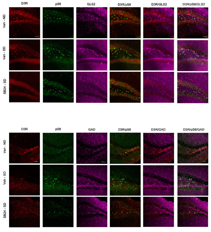
Figure 9.
Representative confocal images showing the dentate gyrus coronal sections immunostained for dopamine type 3 receptor (D3DR, red), phosphorylated S6 protein (activated; green), GLS2 (magenta, glutamatergic neurons), and GAD (magenta, GABAergic neurons) from controls (veh-ND), from mice exposed to a social defeat agonistic encounter after 60 days of abstinence (veh-SD) and from mice receiving an injection of the D3DR antagonist SB-277011-A (24 mg/kg i.p.) prior to the social-defeated agonistic encounter after 60 days of abstinence (SB24-SD). Colocalization of D3DR/GLS2, pS6/GLS2, DRD3/pS6/GLS2, D3DR/GAD, pS6/GAD, and DRD3/pS6/GAD are also shown in the figure. Scale bars: 50 μm.

