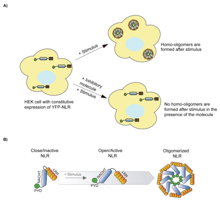
Figure 3.
Schematic illustration showing the formation and the inhibition of NLR oligomers. (A) Illustration of NLR homo-oligomers formation after a stimulus treatment and the inhibition of homo-oligomers formation in the presence of an inhibitor. If the NLR sensor is fused to a fluorescent reporter protein, the formation of homo-oligomers appears as a puncta across the cytoplasm of the cell. If the cell treatment directly activates the NLR inflammasome, this technique is a reliable screening method to identify specific inflammasome inhibitors. If the signaling leading to inflammasome activation requires different steps, positive compounds could either directly block the inflammasome, or any of these steps. (B) Diagram of possible conformational changes of a closed/inactive NLR to an open/active NLR that favor the formation of homo-oligomers.

