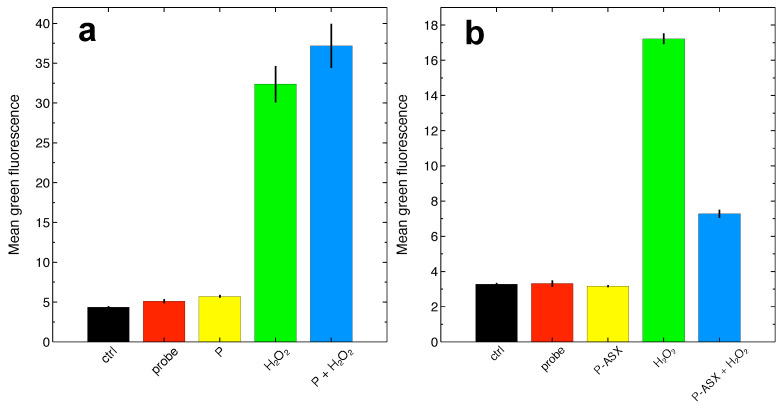
Figure 4.
Determination of ROS levels at the single cell level by flow cytometry using the fluorescent probe DCF-DA.(a) Experiments carried out with cells treated with empty microparticles (P). Overall, the reported differences are statistically significant (, , ANOVA test). HO significantly increases intracellular ROS with respect to controls, but ROS levels in cells treated with HO and empty particles were not statistically different from cells treated with HO alone, suggesting that, within the power of the measurement, empty particles cannot reverse intracellular HO-induced ROS accumulation (, Tukey-HSD post-hoc test). (b) Experiments carried out with ASX-loaded microparticles (P-ASX). The reported differences are statistically significant (, , ANOVA test). ASX-loaded microparticles can significantly reduce intracellular HO-induced ROS accumulation (, Tukey-HSD post-hoc test). In both panels, the labels refer to: ctrl, cell autofluorescence; probe, fluorescence measured with cells loaded with the DCF-DA probe; P or P-ASX, fluorescence measured from cells loaded with DCF-DA and treated with empty (P) or ASX-loaded (P-ASX) microparticles; HO, fluorescence measured from cells loaded with DCF-DA and treated with HO; P or P-ASX + HO, fluorescence measured from cells loaded with DCF-DA, treated with HO and with empty (P) or ASX-loaded (P-ASX) microparticles.

