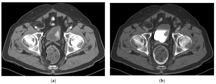
Figure 3.
CT of a bladder mass during the urothelial phase and excretory phase (a) Axial CT image obtained during the urothelial phase shows a hyperenhancing bladder mass. Bladder tumors tend to be hypervascular and scanning the pelvis during the urothelial phase on CT may aid in tumor evaluation. (b) Axial CT image obtained during the excretory phase shows the same mass as a filling defect surrounded by excreted urine in the bladder.

