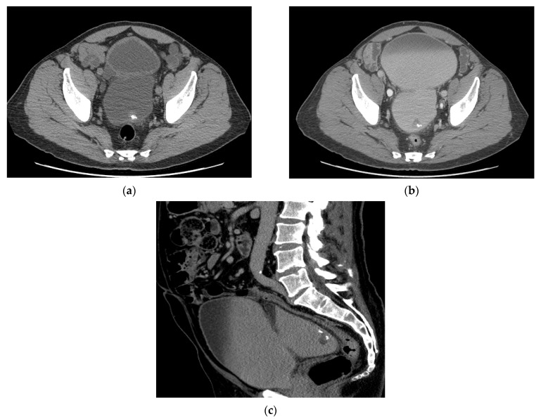
Figure 4.
CT urogram with partially calcified urothelial carcinoma within a large bladder diverticulum (a) Axial CT image obtained without contrast shows a large bladder diverticulum containing a partially calcified lesion along the posterior aspect representing urothelial carcinoma. (b) Axial CT image obtained during the excretory phase and (c) Sagittal excretory phase image show the same mass as a filling defect surrounded by excreted urine in the bladder.

