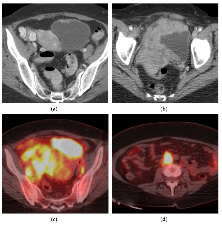
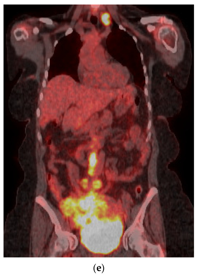
Figure 7.
Marked progression of locally advanced bladder cancer and metastatic disease. (a) Axial CT image demonstrates a small lesion in the anterior bladder which went untreated for 16 months. (b) Follow up axial CT image showing significant progression of the mass which now extends beyond the bladder to involve the uterus, right adnexa, and right pelvic side wall. (c) Axial PET/CT image redemonstrating the locally advanced mass with bilateral pelvic lymphadenopathy. (d) Axial PET/CT image shows metastatic retroperitoneal lymphadenopathy. (e) Coronal PET/CT image also showing metastatic left supraclavicular lymphadenopathy. Biopsy of the left supraclavicular node showed poorly differentiated carcinoma compatible with bladder primary.


