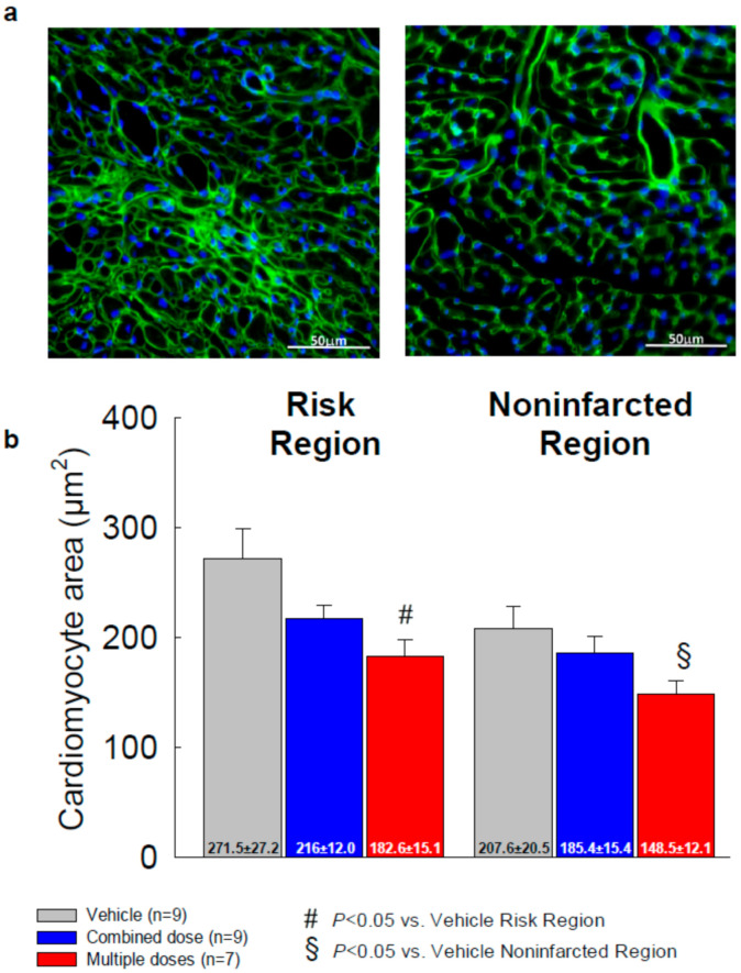
Figure 6.
Analysis of the cross-sectional area of cardiac myocyte (n = 7~9/group). Myocyte cross-sectional area was assessed by immunostaining of cardiac myocytes with DAPI (blue) and WGA (green) to facilitate identification of cell membranes. (a) Representative microscopic images of WGA-stained LV sections; (b) quantitative analysis of myocyte cross-sectional area in the risk and noninfarcted regions. All data are represented as the mean ± SEM.

