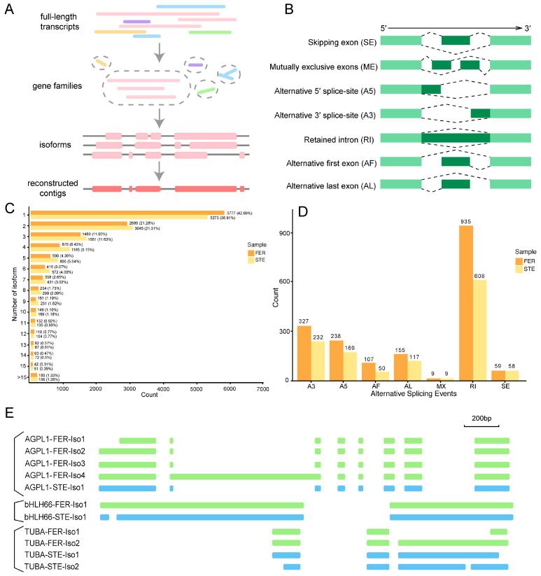
Figure 6.
Comparison of different alternative splicing (AS) among FER and STE. (A) Schematic diagram of Cogent assembly; (B) visualization of seven AS modes; (C) distribution of isoform numbers in FER and STE; (D) different types of AS events in FER and STE; (E) the model graph shows AGPL1/bHLH66/TUBA generating different transcript isoforms of RI detected in two tissues. A3: alternative 3′ splice-site; A5: alternative 5′ splice-site; AF: alternative first exon; AL: alternative last exon; MX: mutually exclusive exons; RI: retained intron; SE: skipping exon.

