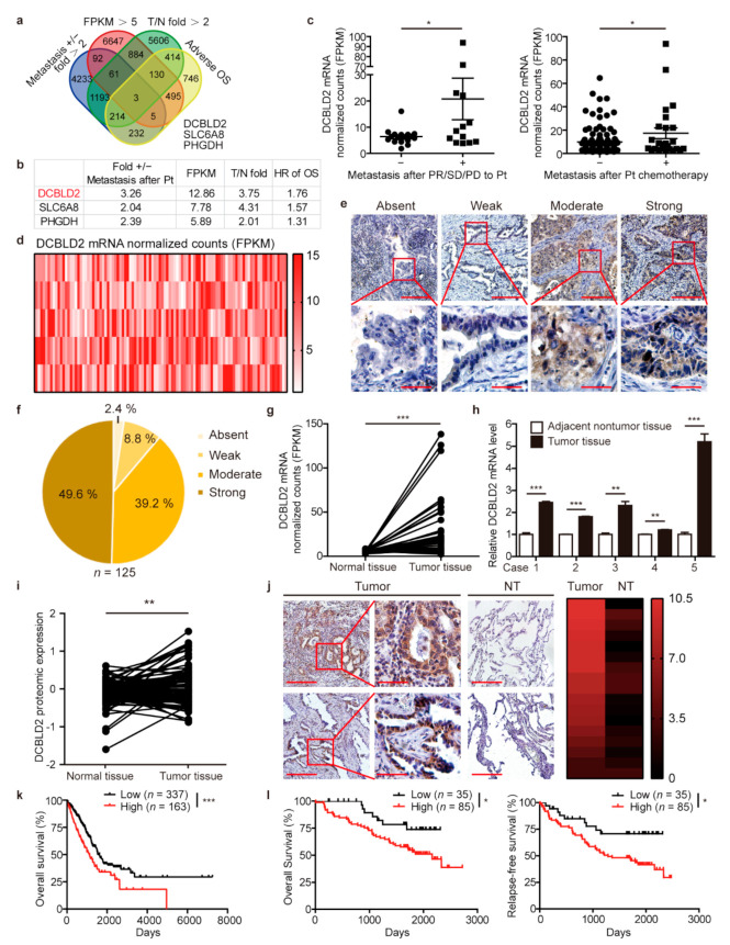
Figure 2.
DCBLD2 is a candidate gene involved in cisplatin-induced metastasis. (a) Screening strategy for key genes related to distant metastasis after platinum-based chemotherapy. (b) Ranking of the 3 identified candidate genes according to the 4 criteria. (c) Differential expression of DCBLD2 at the transcriptional level in LUAD patients from the TCGA database with or without distant metastasis when responding to platinum agents with a partial response, stable disease, or progressive disease (left). DCBLD2 expression in LUAD patients with distant metastasis who received platinum agents (right). * p < 0.05, t-test. (d) The heat map of DCBLD2 expression in mRNA normalized count (log 2) in LUAD tissues from the TCGA database (n = 515). (e,f) Representative images (e) and distribution (f) of DCBLD2 expression in LUAD tissues by IHC assay (n = 125). Scale bar: 200 μm (top), 50 μm (bottom). (g) Transcriptional analysis of the differential expression of DCBLD2 in paired LUAD tissues and normal tissues from the TCGA database (n = 57), p = 0.0003, paired t-test. (h) DCBLD2 expression at the mRNA level in LUAD tissues and adjacent nontumor tissues by RT-PCR (n = 5). ** p < 0.01, *** p < 0.001, t-test. (i) Differential expression of DCBLD2 at the protein level in LUAD tissues and paired normal tissues from the CPTAC database (n = 102), ** p < 0.01, paired t-test. (j) Representative images of DCBLD2 expression in LUAD tissues and paired nontumor tissues (n = 17) by IHC assay. Scale bar: 200 μm (left), 50 μm (middle), 200 μm (right). Wilcoxon’s rank tests, Z = −3.524, p < 0.001. (k) Kaplan-Meier analysis of OS of 500 LUAD patients from the TCGA database stratified by DCBLD2 levels. The cutoff for DCBLD2 expression was 9.5 FPKM. Log-rank tests, p = 0.0002. (l) Kaplan-Meier analysis of OS and RFS of 120 LUAD patients according to different DCBLD2 levels. The cutoff line for H score was 5. Log-rank tests, p = 0.0418 and p = 0.0140, respectively.

