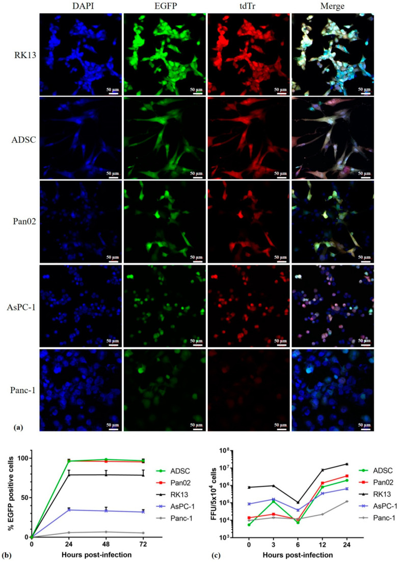
Figure 2.
MYXV infection and replication in pancreatic cancer cells, RK13 and ADSC. Cultures of ADSCs, Pan02, RK13, AsPC-1 and Panc-1 cells were infected: (a) with vMyx-EGFP/tdTr (MOI = 5). At 24 h post infection (p.i.) the infection was visualized by fluorescence microscopy (magn. 20×; scale bar = 50 µm; Zeiss LSM 710 confocal Workstation); blue: DAPI staining (nuclei); green: EGFP fluorescence; red: tdTr fluorescence; (b) with vMyx-EGFP (MOI = 5), collected at the indicated time points and analyzed by means of flow cytometry to determine the percentage of infected EGFP-positive cells; (c) with vMyx-mLIGHT-Fluc/tdTr (MOI = 5) to generate single-step growth curves. Cells were collected at the indicated time points and lysed to determine viral titers. Titers for each sample were performed in triplicate; error bars shown are means ± SD.

