Figure 3.
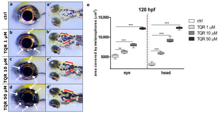
Dose-dependent increase in zebrafish melanophore pigmentation following exposure to tariquidar (TQR) at 120 h post fertilization (hpf). A set of photographs presenting lateral and dorsal views of 120 hpf zebrafish larvae displaying effects of 116 h TQR exposure on the melanophore pigmentation within the eye and dorsal head in four experimental groups: (a,a’) control, (b,b’) exposed to TQR 1 µM, (c,c’) exposed to TQR 10 µM, and (d,d’) exposed to TQR 50 µM. The control larvae presented normal melanophore distribution within both the eye and dorsal head (a,a’). At 120 hpf, the effects of TQR exposure were the continuation of those observed at 72 hpf; however, they were more intense. Progressive hypermelanogenesis was observed with increasing concentration (b,b’;c,c’;d,d’). In the area of the eye, the melanophores began to occupy the area normally covered by the iridophores (b,c,d) (white arrows). Within the dorsal head in the control group, the melanophores were separated and small, while in the TQR exposed groups they were expanded and started to merge creating form closely apposed groups (b’,c’,d’) (red buckle). Additionally, in the control (a’) and 1 µM TQR-exposed group (a’) in the central part of the head, the iridophores were visible (blue arrows), while in 10 µM TQR- and 50 µM TQR-exposed groups these cells were not found. (e) A graph presenting the area covered by melanophores (µm2) measured at 120 h post fertilization (hpf) within the eye and dorsal head (one-way ANOVA/Kruskal–Wallis, GraphPad Prism 5, *** p < 0.001, ** p < 0.01).

