Figure 3.
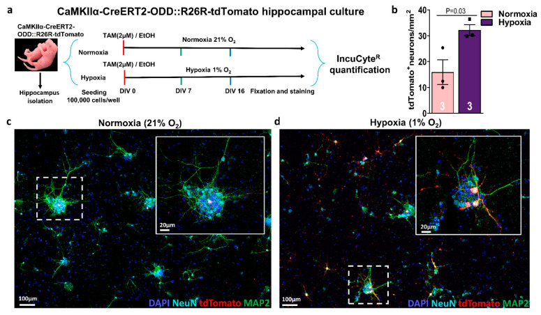
Maturing hippocampal neurons from CaMKIIα-CreERT2-ODD::R26R-tdTomato mice respond to hypoxia also in vitro. (a) Experimental design: Isolation and culture of hippocampal neurons from E17 pups. (Z)-4-hydroxytamoxifen (2 µM) or solvent control (final EtOH concentration always <0.016%) are added to neuron cultures on day in vitro (DIV)0, followed by incubation in the IncuCyteR under either normoxia (21% O2) or hypoxia (1% O2) for seven or 16 days, fixation, staining, and quantification. (b) Quantification of hypoxic neurons, as visualized by tdTomato+ label, reveals an increase under hypoxia at DIV16. (c,d) Representative images of hippocampal neurons stained with neuronal markers NeuN (light blue), MAP2 (green) demonstrate co-localization with tdTomato (red), as shown strongly in hypoxic and less prominent in normoxic conditions. DAPI (blue) was used as a nuclear counterstain. Scale bars represent 100 µm in overview and 20 µm in magnified images. Unpaired Student’s t-test (one-tailed, Welch’s corrections) was used for statistical analysis between conditions; n numbers given in graphs; error bars indicate standard error of mean (SEM).

