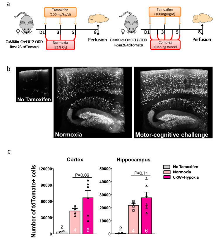
Figure 5.
Light-sheet microscopy (LSM) enables 3D presentation of hypoxic neurons in CaMKIIα-CreERT2-ODD::R26R reporter mice. (a) Experimental design. (b) Illustrative 3D images rendered in maximal intensity modus demonstrate tdTomato+ hypoxic neurons in hippocampus and cortex of female normoxia versus CRW mice; the small image on the left displays a ‘no tamoxifen’ control brain with very few scattered tdTomato+ neurons. Scale: 1.96 × 2.28 × 0.4 mm. (c) LSM quantification of tdTomato+ cells in cortex and hippocampus of eight-week-old female mice show a clear tendency of an equally strong increase under CRW and hypoxia (groups thus pooled) as compared to normoxia, with the ‘no tamoxifen’ condition being negligible. Unpaired Student’s t-test (two-tailed, Welch’s corrections); n numbers given in graphs; error bars indicate standard error of mean (SEM).

