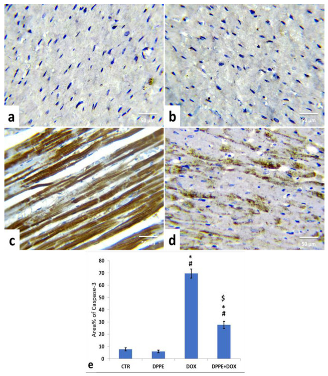
Figure 5.
Immunohistochemical staining of cysteine aspartate specific protease-3 (cleaved caspase-3) in the cardiac cells of the experimental rats (IHC, ×400). A control (a), DPPE-treated (b) DOX-treated (c) and DPP + DOX-treated (d) rats. (e) Quantification of caspase-3 expression, the immunohistochemical staining of cleaved caspase-3 was measured as area percent (%) across 10 different fields/section, n = 7 rat/group. Mean values were statistically different from the CTR (# p < 0.05), DPPE (* p < 0.05), and DOX ($ p < 0.05) groups.

