Figure 1.
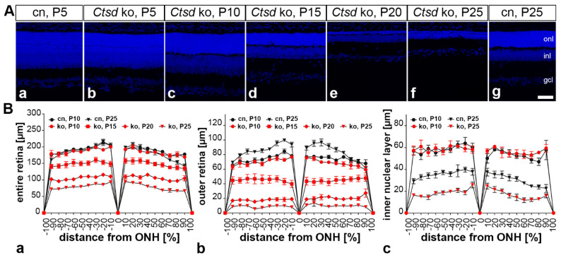
Progressive thinning of the Ctsd ko retina. (A) Retina sections from Ctsd ko mice at different ages (b–f) demonstrate a rapidly progressing retinal dystrophy in the mutant. Retinas from 5- (a) and 25-day-old control mice (g) are shown for comparison. (B) Quantitative analyses revealed no significant thinning of the entire retina (Ba), outer retina (Bb) or inner nuclear layer (Bc) in 10-day-old Ctsd ko mice when compared with age-matched control mice. Significant thinning of the entire retina (Ba), outer retina (Bb) and inner nuclear layer became apparent in 15-day-old Ctsd ko mice and further progressed with increasing age of the mutants (i.e., P20 and P25). For reasons of clarity, only data for selected ages are shown in (B). cn, control; gcl, ganglion cell layer; inl, inner nuclear layer; ko, knock-out; ONH, optic nerve head; onl, outer nuclear layer; P, postnatal day. Scale bar: 50 µm.

