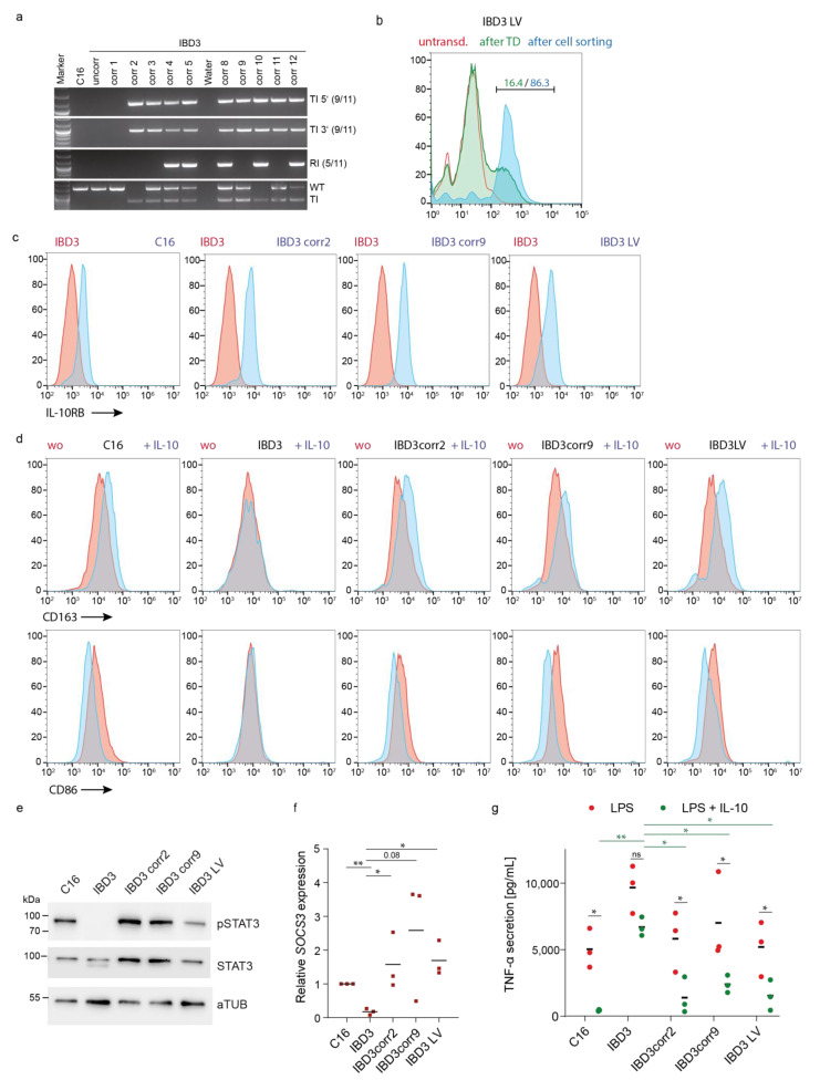
Figure 4.
Genetic correction of iPSCs resulted in functional anti-inflammatory recovery of iPSC-derived macrophages. (a) Genotyping of the donor cassette integration into the AAVS1 genomic locus. PCR analysis using oligonucleotides to detect the right and left flanking arms of the donor cassette in IBD3 subclones to investigate 5′ and 3′ targeted integration (TI). Unspecific random integration (RI) and detection of the physiological wild type (WT) or targeted AAVS1 (TI) locus were used for more detailed validation of the loci. (b) Flow cytometric analysis of IL-10RB surface expression on lentiviral vector transduced IBD3 iPSC (IBD3 LV) before and after purification of transgene expressing cells (red line, untransduced iPSCs; green line, iPSCs after transduction (TD); blue line, sorted iPSCs by FACS). (c) Flow cytometric analysis of IL-10RB expression on corrected and uncorrected myeloid-differentiated suspension cells (red line, IBD3-derived cells; blue line, genetic corrected iPSC-derived cells). (d) IL-10 specific CD163 and CD86 up- or downregulation on uncorrected or corrected iPSC-derived macrophages upon stimulation (red line, without stimulation; blue line, stimulation with IL-10). (e) Western blot analysis of pSTAT3 phosphorylation (pY705, pSTAT3) in iPSC-derived macrophages after IL-10 stimulation. Expression of STAT3 and alpha-Tubulin (aTUB) as controls. (f) Upregulation of SOCS3 mRNA expression upon IL-10 stimulation in iPSC-derived macrophages detected by qRT-PCR (mean of biological replicates, n = 3; * p ≤ 0.05, ** p ≤ 0.01 unpaired t-test). (g) IL-10-mediated regulation of TNF-α secretion in LPS-stimulated uncorrected and corrected iPSC-derived macrophages (mean of biological replicates, n = 3; * p ≤ 0.05, ** p ≤ 0.01 two-way ANOVA and Holm–Šídák multiple comparison test).

