Figure 3.
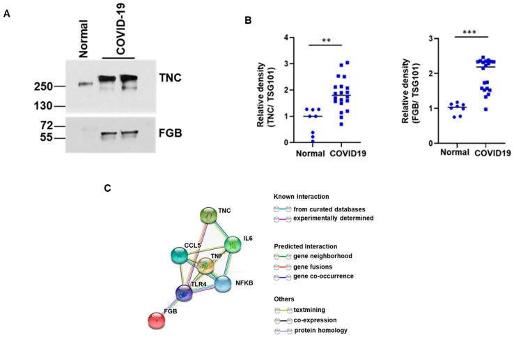
Tenascin-C (TNC) and fibrinogen-β (FGB) are highly present in exosomes of COVID-19 patients. (A) Lysates from COVID-19 plasma exosomes and normal exosomes were subjected to Western blot analysis for TNC and FGB using specific antibodies and a representative image is shown. (B) Dot plots for quantitative Western blot band intensities by densitometry analysis using ImageJ software are shown (n = 8 normal and n = 20 COVID-19 samples). TSG101, an exosomal marker protein, was used for normalization of each sample. (** p < 0.01; *** p < 0.001). (C) String analysis network module represents functional association of TNC and FGB with TLR4/NF-κB signaling. Each node represents all the proteins produced by a single protein coding gene. Colored node represents query proteins and first shell of interactions. Filled node shows 3D structure (known or predicted). Edges represent protein–protein associations for shared function.

