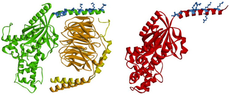
Figure 2.
Localization of positively charged amino acid residues (blue balls and sticks) of the N-terminal fragment in the context of the tertiary and quaternary structure of Gαsβ1γ2 heterotrimer and Gαi1 subunit (red). The Gαs is colored green; the Gβ1 is orange; the Gγ2–yellow. The diagram was generated using coordinates from the PDB: 6X18 and 6CRK and visualized with BIOVIA Discovery Studio 4.0.

