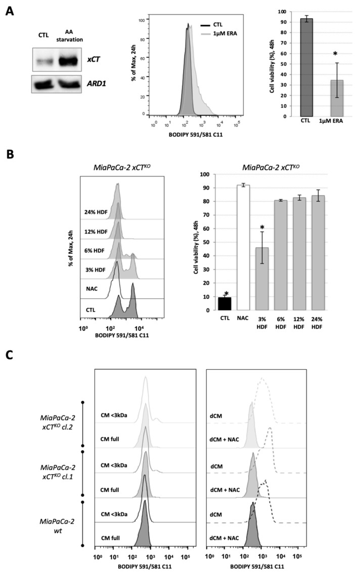
Figure 1.
Fibroblasts reverse the characteristic ferroptosis phenotype of pancreatic ductal adenocarcinoma (PDAC) xCTKO cells. (A). The expression level of xCT in human dermal fibroblasts (HDF) under basal and amino acid (AA) starvation conditions (Western blot, left panel—ARD1 used as loading control), their sensitivity to xCT inhibition by erastin (ERA) seen through the lipid hydroperoxide accumulation after 24 h (BODIPY 591/581 C11 staining, middle panel), i.e., cell viability after 48 h (right bar graph). (B). Optimization of the co-culture conditions: accumulation of the lipid hydroperoxides (left) and cell viability (right) of MiaPaCa-2 xCTKO cells co-cultured with 3%, 6%, 12% or 25%, of HDF cells during 24 h and 48 h, respectively. (C). BODIPY 591/581 C11 staining of MiaPaCa-2 wild type (wt) and xCTKO cells cultured for 24 h in full or fractionated (lower (<) or higher (>) then 3 kDa, left and right panel, respectively) conditional media (CM) of HDF; dCM = dialyzed conditional media (>3 kDa). All experiments have been performed in triplicate and the representative blots and histograms are shown. Bar graphs show mean ± SEM; n = 3; *, p < 0.05, comparison with control group.

