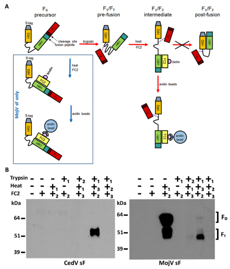
Figure 7.
MojV sF can be triggered to undergo a pre- to post-fusion conformational change. (A) HR2 heptad peptide triggering and capture assay. The henipavirus sF0 is cleaved into the disulfide linked sF1 and F2 subunits by trypsin digestion. The pre-fusion conformation of cleaved sF trimer is triggered into transitioning from pre- to post-fusion conformation by heat application. Upon triggering, pre-fusion sF rearranges into an intermediate structure before reaching its postfusion conformation. This intermediate conformation allows access to the HR1 domain of sF1 by a biotinylated HR2 peptide (FC2). Interaction between intermediate stage sF1 and the FC2 peptide prevents sF from completing its transition into the post-fusion conformation by blocking HR1/HR2 interactions. The FC2 peptide thus “captures” an intermediate conformation of sF as it transitions from pre- to post-fusion conformation. The sF/FC2 peptide complex can then be precipitated by incubation with avidin beads. The dashed line represents the disulfide bond between the F1 and F2 subunits. (B) Soluble MojV F (MojV sF) and CedV F (CedV sF) proteins were treated with various combinations of trypsin, heat and the addition of FC2 peptide. The order of treatment applied in each combination in numbered 1, 2, 3. Treated protein complexes were precipitated with avidin agarose beads, resolved under reducing conditions by SDS-PAGE and analyzed by Western blot. CedV sF was detected with mouse polyclonal anti-CedV sF serum at a 1:1000 dilution. MojV sF was detected with mAb 4G5 at a 1:2500 dilution. The F2 is not detected. This experiment was performed more than 5 times with different preparations of sF glycoprotein trimers.

