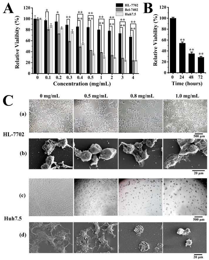
Figure 4.
EPS364 inhibited Huh7.5 cells proliferation and adhesion. (A) Cell viability after different concentrations of the EPS364 (0–4 mg/mL) treatment for 48 h detected by MTT (3-(4,5)-dimethylthiahiazo (-z-y1)-3,5-di-phenytetrazoliumromide) assay. (B) Huh7.5 cell viability after EPS364 treatment (0.5 mg/mL) for different times (0–72 h) detected by the MTT method. (C) The cell morphology after EPS364 treatment for 12 h. (a) HL-7702 cell morphology observed by inverted-phase contrast microscope, (b) HL-7702 cell morphology observed by scanning electron microscope (SEM), (c) Huh7.5 cell morphology observed by inverted-phase contrast microscope and (d) Huh7.5 cell morphology observed by scanning electron microscope (SEM). The cell viability assays were performed in three dependent experiments. * p < 0.05 and ** p < 0.01 versus control.

