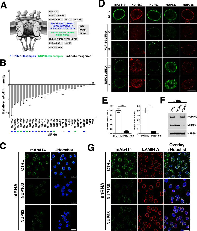
Figure 1. NUP93 and NUP160 nucleoporins are critical for NPC assembly.
A, Schematic illustration of nuclear pore complexes. Subcomplexes are shown in gray boxes. Blue and green nucleoporins are part of NPC scaffold subcomplexes. Asterisk shows the four different FXFG-repeat-containing nucleoporins recognized by the mAb414 antibody. B, Nucleoporins were downregulated in A375 cells using specific siRNA pools (four siRNAs per target) and NPC intensity at the nuclear envelope was quantified 48 hours post-transfection using the mAb414 antibody. 500–2000 cells were analyzed for each well and siRNA treatment was done in duplicate wells. Green and blue dots indicate scaffold nucleoporins and asterisks indicate mAb414-recognized nucleoporins as shown in (A). C, NUP93 and NUP160 were downregulated for 72 hours using the specific siRNA pools described in (B) and NPCs were analyzed by immunofluorescence with the mAb414 antibody. Scale bar 25 μm. D, A375 cells treated with control siRNAs or two different siRNAs against NUP93 and NUP160 for 72 hours were stained against different NPC components. Images show the nuclear cross-sections. Scale bar, 10 μm. E, F, A375 cells stably expressing doxycycline-inducible control shRNAs or shRNAs against NUP93 and NUP160 were treated with doxycycline for 72 hours and the expression levels of both nucleoporins was analyzed by qPCR (E) and western blot (F). G, Immunofluorescence analysis of LAMIN A and mAb414 in A375 cells after 72 hours of Control, NUP160, or NUP93 shRNA induction. Scale bar 25 μm. Data are mean ± s.d. Experiments are representative of 3–5 independent repeats.

