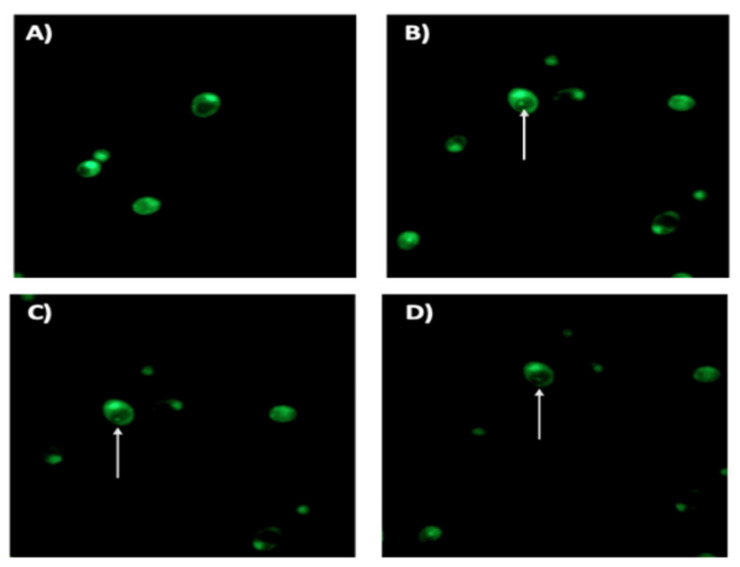
Figure 4.
Fluorescence microscopy of a 48 h H. pylori J99-C. albicans ATCC 10231 co-culture in the presence of ¼ MIC AMX showing yeast cells containing BLBs, putatively H. pylori. (A) shows a pure culture of C. albicans 10231 (negative control) while (B–D) show micrographs of a viable BLB (arrow) within a viable yeast cell taken at 1 s intervals. AMX: amoxicillin, BLBs: bacteria-like body.

