Figure 5.
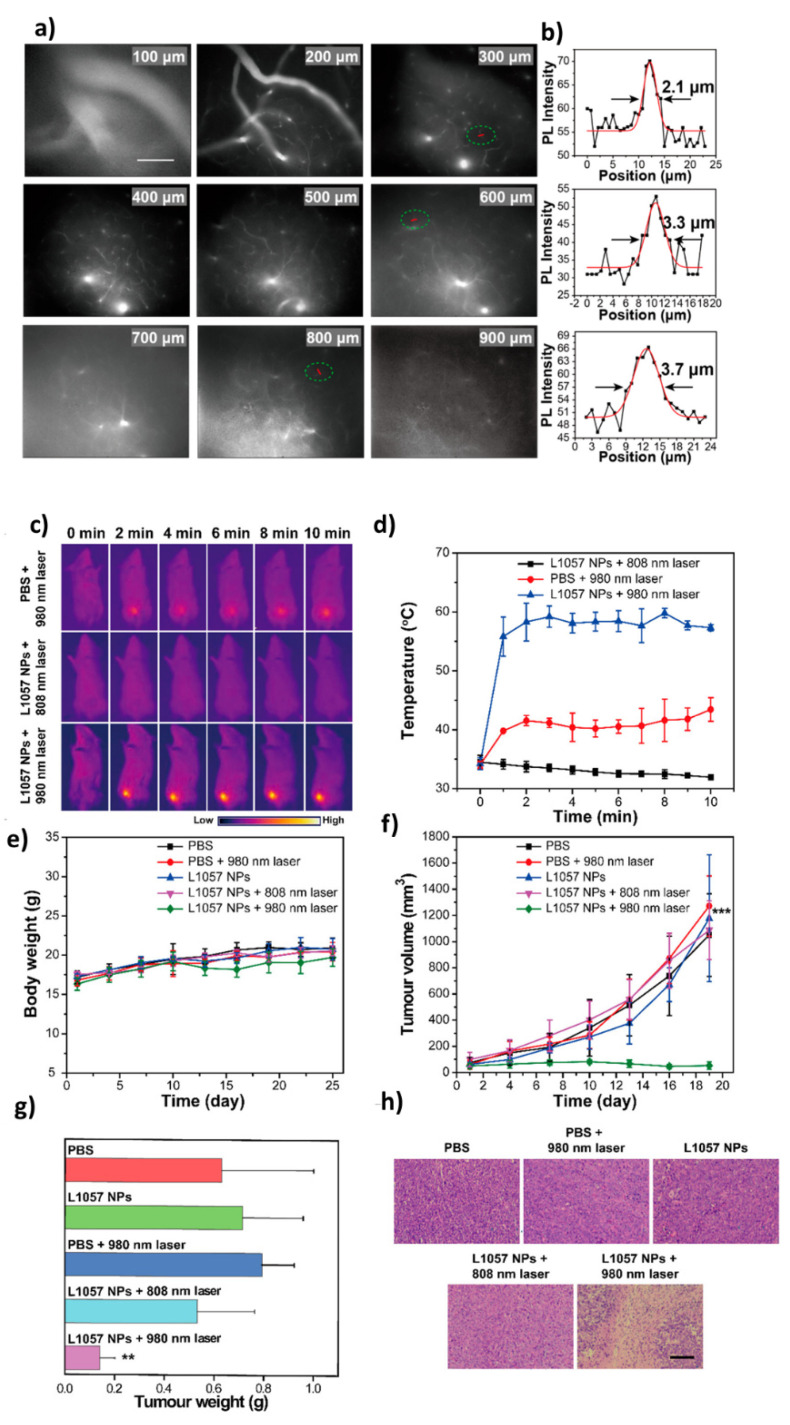
Real-time in-vivo NIR-II fluorescence microscopic imaging of mouse brain vasculature. (a) Cerebrovascular imaging at various depths (100–900 μm) after the intravenous injection of L1057 NPs. The excitation wavelength was 980 nm. Scale bar: 100 μm. (b) Cross-sectional fluorescence intensity profiles (and Gaussian fits (red) with fwhm indicated by arrows) along the red lines circled with green in panel a; PTT efficacy of L1057 NPs on tumors. (c,d) PTT images (c) and corresponding temperature changes (d) of 4T1-tumor-bearing mice under irradiation with an 808 (0.33 W/cm2) or 980 nm (0.72 W/cm2) laser. (e,f) Body weight (e) and tumor volume (f) curves of tumor-bearing mice at different time points after receiving PTT. (g,h) Tumor weight (g) and H&E staining (h) of the tumor tissues from mice sacrificed at day 18 post-PTT treatment. Scale bar: 100 μm. Results are presented as the mean ± S.D., n = 5. Statistical significance was calculated using one-way ANOVA with the Tukey posthoc test. *** p < 0.001. (Reprinted Permission from American Chemical Society, 2020) [61].

