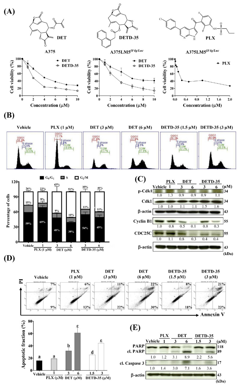
Figure 2.
DET and DETD-35 inhibited melanoma cell proliferation and induced G2/M cell cycle arrest and apoptosis in A375LM5IF4g/Luc lung-seeking melanoma cells. (A) Cell viability was determined by MTT assay. A375 and A375LM5IF4g/Luc cells were incubated with vehicle, DET or DETD-35 for 24 h and vehicle or PLX for 72 h. (B) A375LM5IF4g/Luc cells were incubated with vehicle, DET, DETD-35 or PLX for 24 h. The cells were collected, fixed with ethanol, and stained with propidium iodide, and DNA distribution in the cells was analyzed by flow cytometry. Top, DNA content in the cell. Bottom, DNA percentage in G0/G1, S and G2/M stage. Data are mean ± SD, n = 3. (C) A375LM5IF4g/Luc cells were incubated with the same agents as in (B) for 24 h and the expression of the cell cycle proteins were analyzed using Western blotting. Increased/decreased protein levels among treatments are presented as a fold change to the vehicle control after normalization to β-actin. (D) A375LM5IF4g/Luc cells were incubated with vehicle, DET, DETD-35 or PLX for 48 h. The apoptotic population was determined by Annexin V/PI staining and flow cytometry. Top, related quadrant diagrams showing apoptotic cell status. Bottom, quantitative data are mean ± SD, n = 3. Means with significant differences are denoted with different letters (one-way ANOVA, p ≤ 0.05). (E) A375LM5IF4g/Luc cells were incubated with the same agents as in (D) for 48 h and the expression of the apoptotic proteins were analyzed by Western blotting. Increased/decreased protein levels among treatments are presented as a fold change to the vehicle control after normalization to β-actin.

