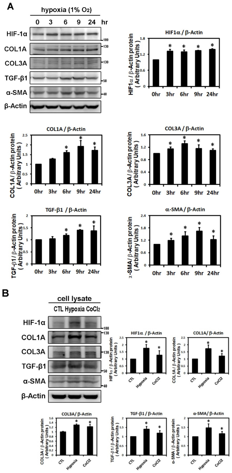
Figure 1.
Effects of hypoxic treatment on the expression of HIF-1α and profibrotic proteins. Expression of the indicated proteins in cellular homogenates was measured by Western blot. Each lane contained a total of 25 μg of protein and the blots were probed with the indicated antibody. All data were normalized to the β-actin loading and blotting control. Bar graphs indicate band intensity as determined by densitometry and the fold increase in intensity over control. Panel A: Exposure of cultured HL-1 cells to hypoxia (1% O2) for the indicated times resulted in time-dependently increased protein levels of HIF-1α (maximum reached at 3 h), COL3A (maximum reached at 6 h) and TGF-β1, α-SMA, COL1A (maxima reached at 9 h). Panel B: Representative Western blots and quantitative analysis of protein levels of HIF-1α and fibrosis-related proteins (COL1A, COL3A, TGF-β1, α-SMA) in HL-1 cells treated with hypoxia (1% O2) or CoCl2 (10−4 M) for 6 h. The data are mean ± SEM (n = 4 per group, * p < 0.05 vs. control) from four independent experiments.

