Figure 1.
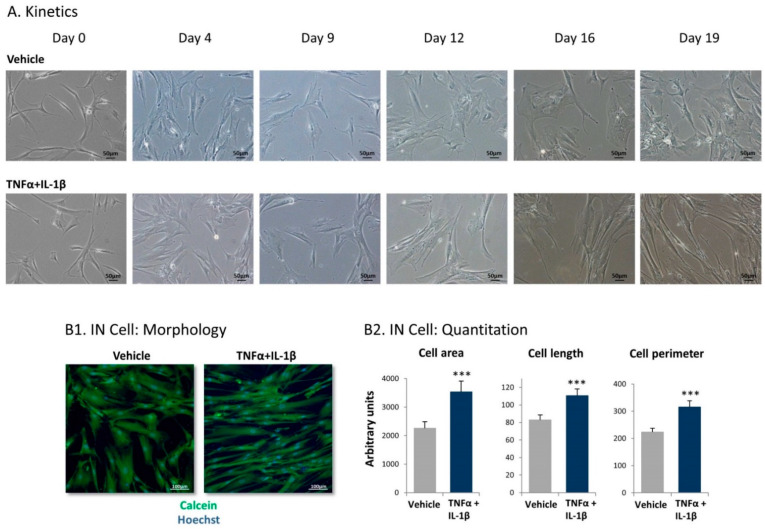
Following persistent stimulation of mesenchymal stem cells (MSCs) by tumor necrosis factor α (TNFα) + interleukin 1β (IL-1β), the resulting cells acquire elongated cancer-associated fibroblast (CAF)-like morphology. Human MSCs were exposed to TNFα (50 ng/mL) + IL-1β (0.5 ng/mL) or to vehicles. Cytokine concentrations were selected based on the considerations described in the Materials and Methods section. (A) At different time points, the cells were photographed by light microscopy. Images from a representative experiment out of n > 3 are presented. Bar, 50 μm. (B) Determination of cell characteristics using IN Cell technology in cells that were treated using the cytokines/vehicles for 18 days and were then subjected to IN Cell analysis. (B1) Cell morphology was detected by calcein (green) and Hoechst (blue) staining. Images of cell morphology from a representative experiment out of n = 3 are presented. Bar, 100 µm. (B2) Quantification of cell characteristics by the IN Cell technology. *** p < 0.001. The results of a representative experiment of n = 3 are presented.

