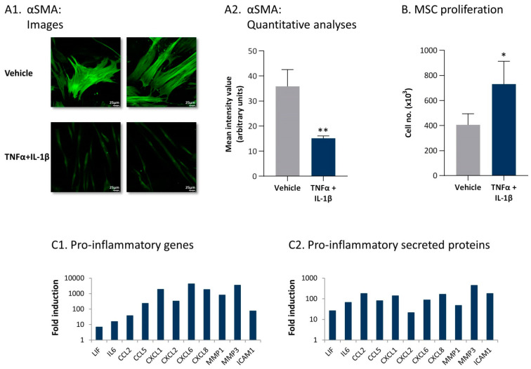
Figure 5.
Persistent stimulation with TNFα + IL-1β leads to the conversion of MSCs to inflammatory CAFs. (A,B) Human MSCs were exposed to persistent TNFα + IL-1β stimulation (concentrations as in Figure 1) or to vehicles, generally for 14–18 days. (A) αSMA expression was determined by confocal analyses. (A1) Two images of each treatment, derived from a representative experiment out of n > 3, are presented. Bar, 25 μm. (A2) Quantitative analyses of the fluorescence intensity performed by the ImageJ program on images of the representative experiment (n ≥ 5 images for each treatment). ** p < 0.01. (B) Cell proliferation was determined by cell counts at the end of the stimulation process. Average ± SD of n > 3 is presented. * p < 0.05. Time-dependent analysis of MSC cell numbers along the stimulation process are demonstrated in Figure S2. (C) Expression of pro-inflammatory genes and secreted proteins, determined at the end of the stimulation process, is demonstrated. (C1) mRNA expression was determined by transcriptome analyses performed at day 14, with three independent biological repeats. padj < 10 × 10−10–padj < 5 × 10−271, depending on the gene. (C2) Expression of secreted proteins, determined by secretome analyses performed at days 18–19 with three independent biological repeats. p < 10−3–p < 7.5 × 10⁻⁷, depending on the protein. Data are presented as the fold induction of values obtained in cytokine-stimulated cells compared to vehicle-treated cells for all genes and proteins. Tables S1–S4 demonstrate the 60 top upregulated or downregulated genes and proteins, obtained following persistent stimulation of the MSCs by TNFα + IL-1β.

