Figure 9.
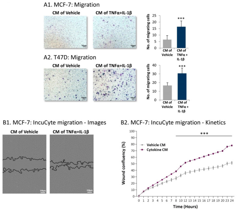
Following persistent stimulation of MSCs with TNFα + IL-1β, the resulting inflammatory CAFs release factors that promote migration of luminal-A BC cells. Human MSCs were exposed to persistent TNFα + IL-1β stimulation (concentrations as in Figure 1) or to vehicles, generally for 14–18 days. Cytokine-devoid CM were collected as described in Figure 7 and were administered to mCherry-expressing MCF-7 and T47D human luminal-A BC cells. (A) Tumor cell migration was determined in fibronectin-coated transwells in response to serum-containing medium. (A1) MCF-7 cells. (A2) T47D cells. Images and quantitative analyses of a representative experiment out of n = 3, for each cell type, are presented. *** p < 0.001. (B) MCF-7 wound closure assay, performed in IncuCyte®. (B1) Representative images taken at 24 h. Bar, 150 µm. (B2) The kinetics graphs of tumor cell migration demonstrate the proportion (%) of the original wound area that became covered by migrating cells at each time point. The results presented are mean ± SEM of 5 replicates for each treatment and are of a representative experiment out of n > 3. *** p < 0.001.

