Figure 3.
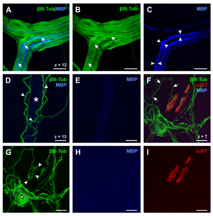
Expression of βIII-Tubulin, MBP, and α-BT in the esophagus (A)–(E): Triple whole mount staining of βIII-tubulin, MBP, and α-BT (α-BT not shown, N° ⑤, Table 2). Axons ((A) and (B), short arrows) of peripheral vagal nerve branches are only partially myelinated (C, arrowheads), while gracile blood vessel-related nerve fibers, which are tightly wrapped around the outer vessel wall, appear βIII-tubulin-positive (D, arrowheads) but always lack myelin since no MBP signal can be detected (E). The lumen of the blood vessel is marked by the asterisk (D). (F–I): Triple whole mount staining of βIII-tubulin, MBP and α-BT (N° ⑤, Table 2) reveals that endplate contacting efferent axons (F, short arrows) are always unmyelinated for a long distance (H) and form a framework in the presynaptic region of the NMJ (G, arrowheads). These findings confirm the results of the ChAT staining protocol (cf. Figure 2E–H). Moreover, βIII-tubulin can also be found in enteric neurons (G, dotted line; nucleus marked by asterisk) of the myenteric plexus and therefore proves to be a suitable neuro-axonal marker for the evaluation of the ENS in the esophagus. ChAT: Cholin acetyltransferase; MBP: Myelin basic protein; Z-step = 1 µm; scale bars 20 µm.

