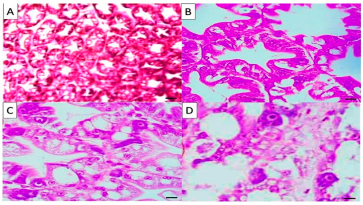
Figure 1.
Hepatopancreas (HP) of Penaeus vannamei: (A) T1 showed normal HP structure and tubule epithelial cells surrounded by hemolytic infiltration; (B) T2 showed slight hemocyte infiltration, and normal hepatopancreas lumen and tubule, (C) T3 showed mild hemocyte infiltration and normal hepatopancreas lumen and tubule; (D) T4 showed highly activation of the hepatic glandular duct system. H&E stain magnification (×200), bar = 50 µm.

