Figure 5.

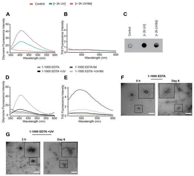
Freshly prepared dGAE samples (100 μM) were incubated without UV (control) or in the presence of UV for 2 h at 4 °C [+2 h UV], and the 2 h UV-exposed sample was further incubated for six days at 37 °C/350 RPM [+2 h UV/6 d]. Measurement of DiY signal over time revealed the induction of DiY by the UV exposed sample (A). ThS fluorescence assay showed no fluorescence intensity indicating there was no assembly in any of the samples (B). Immunoblotting using the T22 antibody, suggested the presence of a large quantity of tau oligomers in the UV-exposed samples compared to control (C). Freshly prepared 1-1000 EDTA samples were incubated without UV for 2 h or 6 days at 37 °C/350 RPM (1-1000 EDTA 6 d) and with UV for 2 h (1-1000 EDTA +UV) or 2 h UV followed by 6 days incubation at 37 °C/350 RPM (1-1000 EDTA +UV/6 d). DiY signal was collected at each time point (D), followed by ThS fluorescence assay, which suggested assembly in the 1-1000 EDTA 6 d sample and not the others (E). TEM imaging at 0 h revealed small, round assemblies in the 0 h [1-1000 EDTA] sample, which assembled into long mature fibrils after 6 days incubation at 37 °C/350 RPM (F). The 1-1000 EDTA + UV samples revealed both small and large clumped assemblies, which remained largely unchanged even after 6 days of incubation at 37 °C/350 RPM. Scale bar, 500 nm (G).
