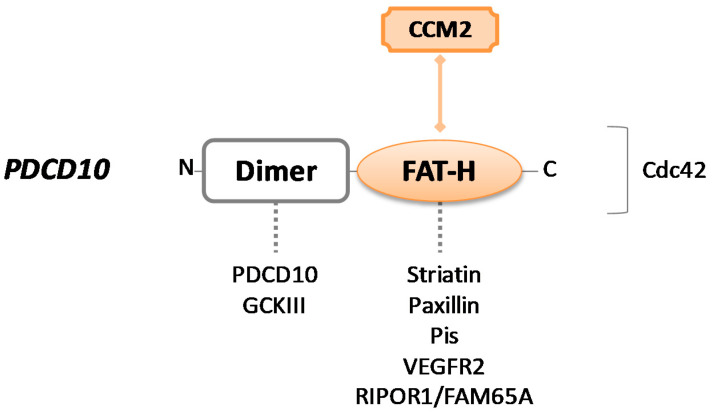
Figure 3.
Schematic representation of CCM3/PDCD10 protein. PDCD10 consists of a dimer site and a focal adhesion targeting homology (FAT–H) domain. Colored forms and lines in the figure indicate the crucial interaction with CCM2 to build the CCM trimeric complex. Dotted lines indicate intermolecular interactions that may occur through each protein domain. The binding site for the Cdc42 protein is unknown.

