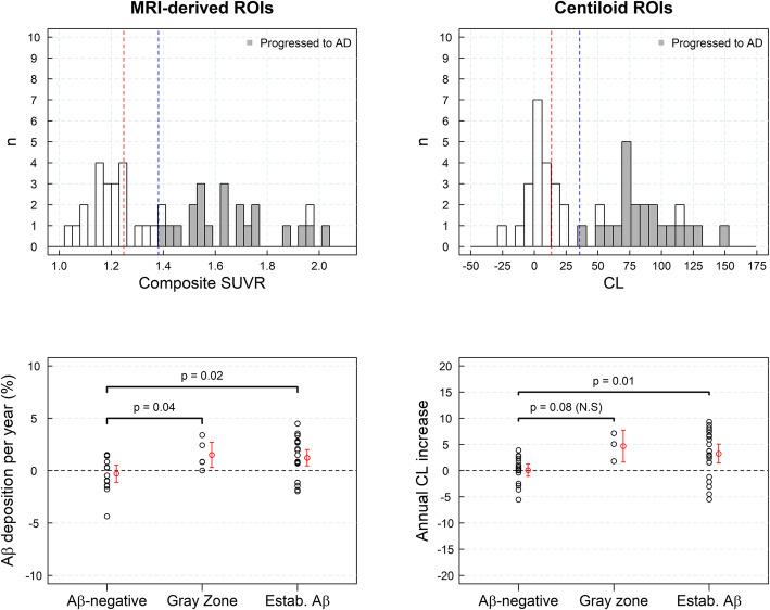
Fig. 5.
Histograms of composite standardized uptake value ratios (SUVRs) and centiloids (CLs) for the sample of mild cognitive impairment (MCI) (n = 44, dataset #4) subjects are shown on the top row. Subjects that progressed to Alzheimer’s disease (AD) dementia after a 4-year clinical follow-up are shown in gray. Red and blue lines represent the SUVR abnormality cutoffs for early Aβ detection and established Aβ pathology, respectively. The rate of Aβ accumulation in MCI subjects (and 95% confidence interval in red) in three categories of the composite SUVR continuum: Aβ-negative, gray zone, and with established Aβ deposition, is shown on the bottom row. ROI region of interest

