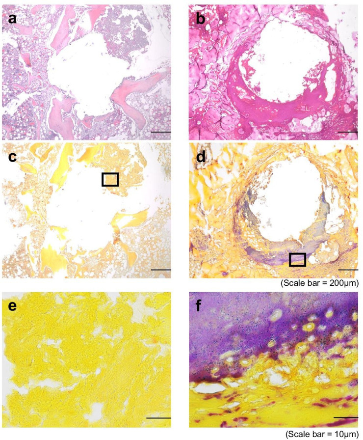
Fig. 5.

Histological evidence of osteomyelitis. Serial sections from a representative sample in the (a, c, e) control aseptic screw group and (b, d, f) chronic periprosthetic joint infection (PJI) group were stained either with haematoxylin and eosin (H&E) (a, b) and imaged at 40× (scale bar = 200 μm), or with Gram’s stain (c-f) imaged at 40× (c, d; scale bar = 200 μm) or 100× (e,f; scale bar = 10 μm).
