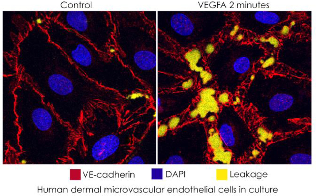
Figure I box 1.
compares control and VEGFA-stimulated microvascular endothelial cells, where leakage is detected by fluorescent streptavidin (yellow) bound to biotin in the substrate, in an in vitro vascular permeability imaging assay (Merck KGaA, Darmstadt). Separations of junctions in cultured endothelial cells exposed to VEGFA are larger and more numerous than occur in vivo.

