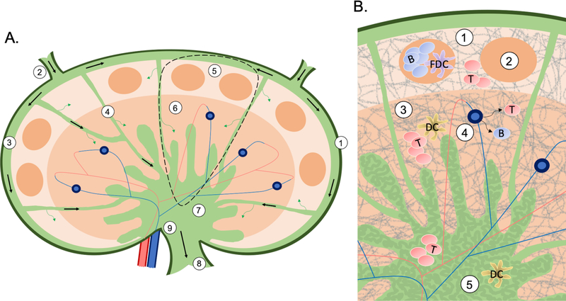
Figure 1:
A) An overview of the structure and flow (arrows) in a lymph node. The capsule (1) is the outmost later of endothelial cells. Lymphatic fluid flows into the node through the afferent (2) lymphatics, around the node through the subcapsular sinus (3), then between each lymph node lobule (dashed region, expanded in B) through the transverse sinuses (4). From the subcapsular sinus fluid can diffuse into the cortex (5), and from the transverse sinuses into the paracortex (6). Finally, lymphatic fluid collects in the medulla (7), and makes its way out of the node through the efferent lymphatics (8) in the hilus (9). B) An overview of the structure and cellular organization of a lymph node lobule. The cortex (1) is the outermost region containing follicles (2), which are made up primarily of B cells and FDCs. The next region is the paracortex (3), containing primarily T cells and DCs, as well as the HEVs (4). Lastly, the medulla (5) is an area of lymph flow and cell trafficking, and its appearance is characterized by a maze-like appearance of medullary cords (dark green) sinuses (light green). Throughout the lymph node the FRC network (gray lines) gives the tissue shape and structure.

