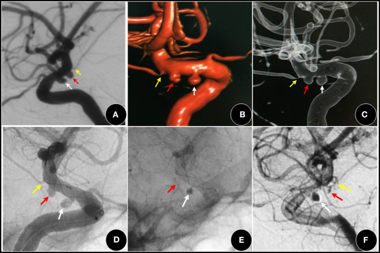
Figure 4.
A 52-year-old female, with a three-month history of dizziness. (A–C) showing a I type aneurysm (red arrow) and an adjacent PCA aneurysm (white arrow). (D–F) two aneurysms were covered by an FD (Tubridge stent), the AChoA aneurysm was barely visible (red arrow). The PCA aneurysm (white arrow) showed obvious retention of contrast media, while the AChoA (yellow arrow) was patent.

