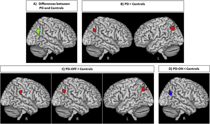
Fig. 2 –
A) Convergence of aberrant (green) and B) convergence of increased (red) resting-state fMRI findings in patients with Parkinson’s disease compared to healthy controls in bilateral parietal lobule. C) Convergence of increased (red) resting-state fMRI findings in patients with Parkinson’s disease in the OFF-state compared to healthy controls in the right supramarginal gyrus and bilateral parietal lobule; D) Convergence of reduced (blue) resting-state fMRI findings in patients with Parkinson’s disease in the ON-state compared to healthy controls in the right parietal lobule. All activations are significant at p < .05 corrected for multiple comparisons using the family-wise error rate in cluster level (cFWE).

