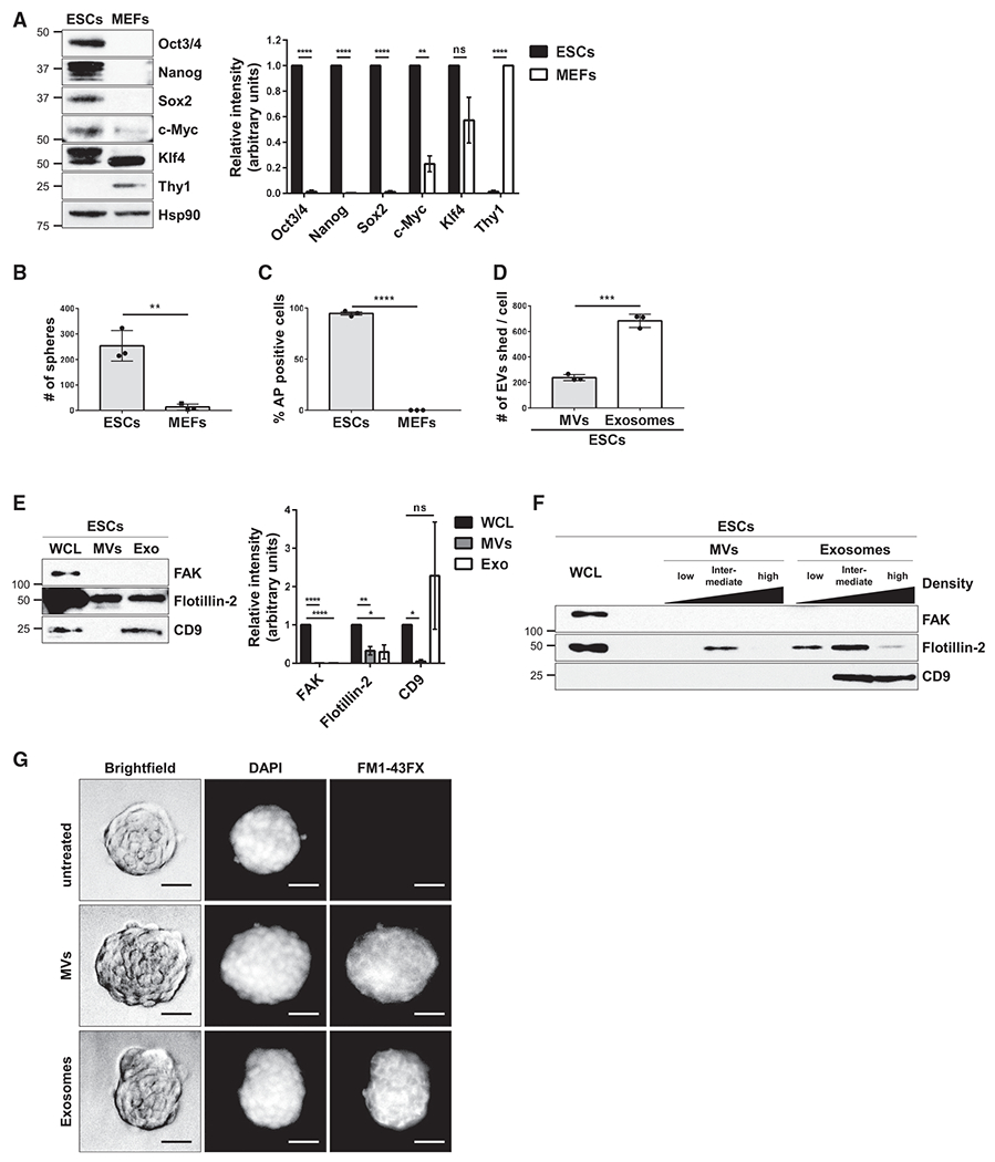
Figure 1. ESCs Generate EVs.

(A) ESCs and MEFs were immunoblotted for markers of pluripotency (i.e., Oct3/4, Nanog, Sox2, c-Myc, and Klf4), the fibroblast marker Thy1, and heat shock protein 90 (Hsp90) as the loading control.*
(B) Sphere formation assays were performed on ESCs and MEFs. See corresponding images in Figure S1C.*
(C) AP activity assays were performed on ESCs and MEFs. See corresponding images in Figure S1E.*
(D) The number of MVs and exosomes released per ESC were determined using nanoparticle tracking analysis (NTA).*
(E) ESCs (whole cell lysate; WCL), MVs, and exosomes (Exo) were immunoblotted for the cytosolic protein FAK, the general EV marker Flotillin-2, and the exosome-specific marker CD9.*
(F) MVs and exosomes isolated from ESCs were further subjected to sucrose density gradient ultracentrifugation. ESCs (WCL) and the resulting fractions were immunoblotted for the same proteins described in (E).
(G) MVs and exosomes from ESCs that had been labeled with the fluorescent membrane dye FM1-43FX were incubated with cultures of ESCs for 1 h, at which point the cells were washed extensively, fixed, and stained with DAPI to label nuclei. Brightfield and fluorescent microscopy images of the assay are shown. Scale bar, 50 μm.
*The data shown in (A)–(E) are presented as mean ± SD. All experiments were performed at least three independent times, and statistical significance was determined using Student’s t test; ****p < 0.0001; ***p < 0.001; **p < 0.01; *p < 0.05; and ns, not significant. See also Figure S1.
