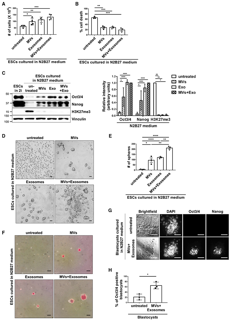
Figure 3. EVs from ESCs Promote Stem-Cell-Related Phenotypes.

(A) Cell growth assays were performed on ESCs cultured in N2B27 medium supplemented without (untreated) or with MVs and/or exosomes from ESCs.*
(B) Cell death assays were performed on ESCs cultured in N2B27 medium supplemented without (untreated) or with MVs and/or exosomes from ESCs.*
(C) ESCs cultured in 2i+LIF medium (lane 1) or N2B27 medium supplemented without (untreated) or with MVs and/or exosomes from ESCs (lanes 2–5) were immunoblotted for Oct3/4, Nanog, H3K27me3, and vinculin as the loading control.*
(D) Images of sphere formation assays performed on ESCs cultured in N2B27 medium supplemented without (untreated) or with MVs and/or exosomes from pluripotent ESCs. Scale bar, 100 μm.
(E) Quantification of the assays shown in (D).*
(F) Images of AP activity assays performed on ESCs cultured in N2B27 medium supplemented without (untreated) or with MVs and/or exosomes from ESCs. Cells positive for AP activity are red. Scale bar, 100 μm.
(G) Brightfield and fluorescent microscopy images of blastocysts isolated from pregnant mice cultured in N2B27 medium supplemented without (untreated) or with MVs and exosomes from ESCs. The blastocysts were stained for Oct3/4 and Nanog, and DAPI was used to label nuclei. Scale bar, 50 μm.
(H) Quantification of Oct3/4-positive blastocysts for each condition shown in (G). This assay was performed three separate times, with a minimum of 13 blastocysts being evaluated per condition in each assay, before the results were averaged and plotted.*
*The data shown in (A)–(C), (E), and (H) are presented as mean ± SD. All experiments were performed at least three independent times, and statistical significance was determined using Student’s t test; ****p < 0.0001; ***p < 0.001; **p < 0.01; and *p < 0.05. See also Figure S3.
