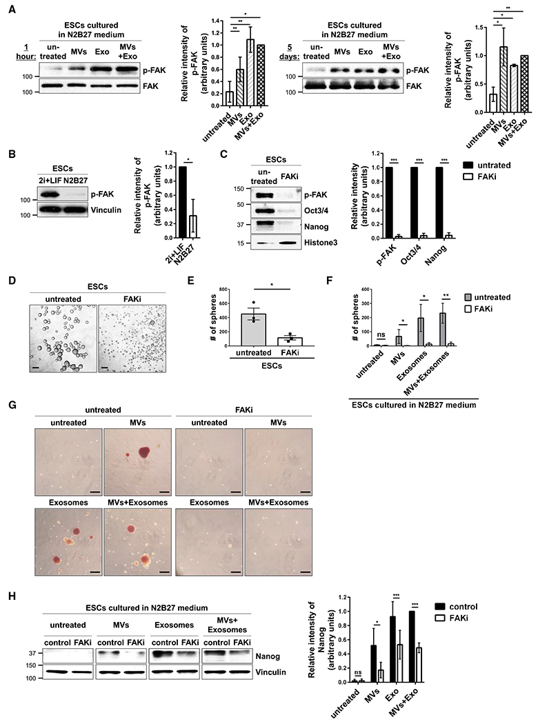
Figure 5. EVs from ESCs Help Maintain Stemness by Activating FAK in Recipient Cells.

(A) ESCs cultured in N2B27 medium supplemented without (untreated) or with MVs and/or exosomes from ESCs for 1 h (left) and 5 days (right) were immunoblotted for phosphorylated FAK (p-FAK) and total FAK.*
(B) ESCs cultured in 2i+LIF or N2B27 medium were immunoblotted for phosphorylated FAK (p-FAK) and vinculin.*
(C) ESCs cultured in 2i+LIF medium supplemented without (untreated) or with 7.5 μM FAK inhibitor III (FAKi) were immunoblotted for phosphorylated FAK (p-FAK), Oct3/4, Nanog, and histone 3 as the loading control.*
(D) Images of sphere formation assays performed on ESCs maintained in 2i+LIF medium supplemented without (untreated) or with 7.5 μM FAK inhibitor III (FAKi). Scale bar, 100 μm.
(E) Quantification of the assays shown in (D).*
(F) Quantification of sphere formation assays performed on ESCs cultured in N2B27 medium supplemented with MVs and/or exosomes produced by ESCs and treated without (untreated, shaded bars) or with 7.5 μM FAK inhibitor III (FAKi, clear bars).*
(G) Images of AP activity assays performed on ESCs cultured in N2B27 medium supplemented with MVs and/or exosomes from ESCs and treated without (untreated) or with 7.5 μM FAK inhibitor III (FAKi). Scale bar, 100 μm.
(H) ESCs cultured in N2B27 medium supplemented with MVs and/or exosomes from ESCs and treated without (control) or with 7.5 μM FAK inhibitor III (FAKi) were immunoblotted for Nanog and vinculin as the loading control.*
*The data shown in (A)–(C), (E), (F), and (H) are presented as mean ± SD. All experiments were performed at least three independent times, and statistical significance was determined using Student’s t test; ***p < 0.001; **p < 0.01; *p < 0.05; and ns, not significant. See Figures S4–S6.
