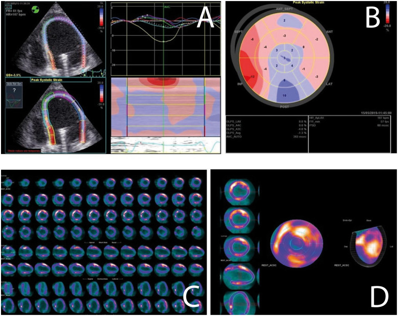
Figure 6.
Cardiac viability assessment with different imaging techniques in the same patient while on Impella support. (A and B) Speckle tracking echocardiography analysis presented as waveform (A) and bull’s eye plot (B). (C and D) Cardiac positron emission tomography analysis similarly presented as short- and long-axis slices (C) and bull’s eye plot (D).

